3D printers print functional prostheses made of polymers and metals. 3D bioprinters print bones, joints, tissues, and even organs from living cells. This has already changed medicine. Large companies order the printed liver tissue to speed up the study of medicine. Scientists are preparing to print out the first human organ. This breakthrough is planned for 2030, but in the meantime, it is easier to find a 3D printer in dentistry.
Why is 3D printing developing so fast in this field of medicine? The explanation is on the surface-problems with teeth happen to all people and no one ignores treatment. Because it hurts. Therefore, the introduction of innovations in dentistry will pay off faster than in Oncology, for example. Another reason is that replacing teeth is easier than replacing bones and organs. No need for surgery — just open your mouth wider and all your teeth are freely accessible.
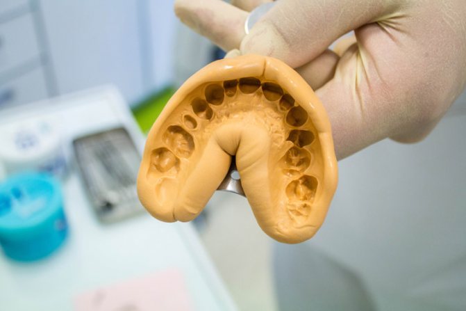
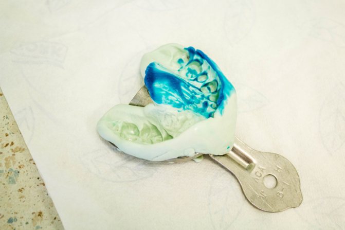
Dental technicians use gypsum and elastic polymers to create a dental impression. This process takes place in several stages and requires constant correction of the impression. The impression itself holds its shape for a limited time, then it is deformed and then it must be made again.
For 3D printing, the patient’s teeth are modeled together with the jaw in a 3D editor. If you need a complete replacement of the jaw, then you need to model the entire oral cavity — this is much easier to do in a 3D editor. Here the model can be divided into individual elements of any size, and its entire shape is easier to control. The 3D printer immediately prints the 3D model with polymers and metal, which speeds up treatment, saves on material and tools for casts.
3D printer for dentistry eliminates the need for manual modeling
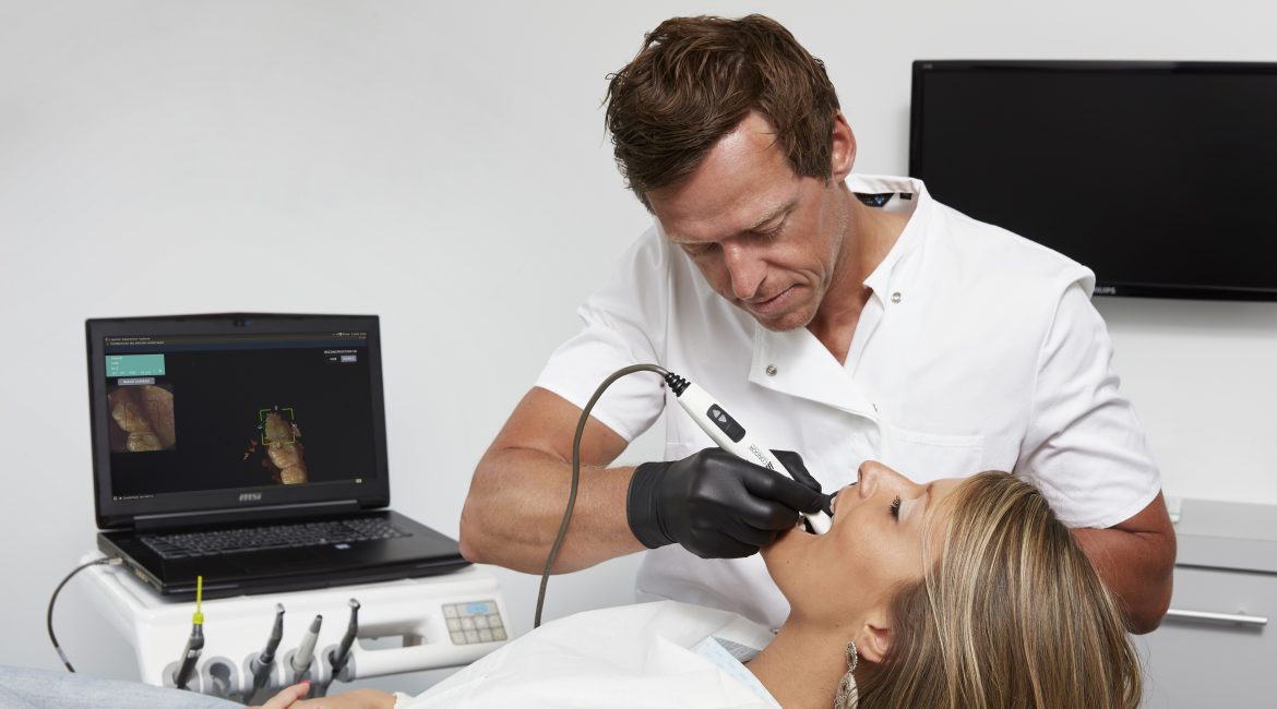
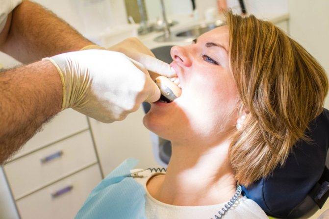
crowns, dentures, and other products. Clients of dental clinics do not wait for the installation of the final design. going through several stages of refinement and fitting. 3D scanning of the oral cavity provides accurate parameters for 3D modeling of the crown or jaw.
Images obtained from three dimensional scanning of the oral cavity are used for building 3D models:
crowns’;
implants’;
plaster model;
bridges;
unique orthodontic tools.
Table of Contents
3D printing technologies in dentistry
And 3D model printing two main printing technologies are used in dentistry:
- selective laser sintering-SLS;
- selective laser melting-SLM.
- layer-by-layer application of quick-drying polymers-WDM.
- SLS uses a laser to selectively sinter layers of metal powder.
The laser in SLM melts the layers, providing less porosity of the metal of the product.
The source material for SLM printing is fine powder
based on a metal alloy. The laser beam melts the powder particles, connecting them together. The next layer of the alloy is applied to the created layer, then another one, and as a result, the finished product of the desired volume and shape is obtained.
3D printers allow you to print complex structures and shapes of prostheses
directly from your computer. Titanium and its alloys, chromium, and cobalt are also used for the manufacture of prostheses. WDM printers layer the quick-drying polymer in layers that are firmly fused until the polymer loses its viscosity.
Three-dimensional printing technologies-melting, sintering, partial or complete melting of the material, etc. make it possible to create a solid structure made of polymers and metals.
Cost of 3D dental prosthetics
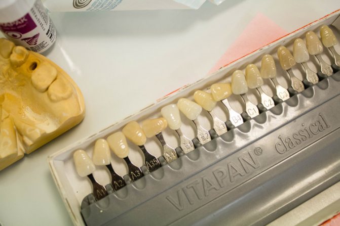
3D prosthetics is also famous for the high aesthetics of the prostheses made with it. The materials used are hypoallergenic, have a wide range of shades, and exactly repeat your own teeth-in color, transparency and Shine.
The cost of 3D prosthetics practically does not differ from the classic method of manufacturing ceramic and zirconium prostheses, since payment for the work of the dental laboratory is completely excluded from the production process. So if you have been thinking about installing prosthetics of high aesthetics for a long time, then 3D prosthetics is an option that is really worth considering as an alternative to traditional methods.
3D printer for dentistry: price and models
Special medical SLM and WDM 3D printers are used in dentistry. They lay the polymer layer by layer, which becomes solid after drying.
Three-dimensional printers for dental purposes
cost ~$20,000.
Some companies, like Stratasys, do not disclose the price, offering to contact and discuss the cost through the site. The following printer models are available on the market:
As you know, medicine has been undergoing significant changes in recent years, which allows patients to get all their guarantees regarding safety, efficiency, reliability, comfort and a good result of treatment. Such positive trends also occur in the dental industry, and in particular in dental implants.
Today, everyone can safely decide to transform their smile with the help of 3D implantation, or rather, thanks to 3D technologies in dentistry. In the article below, we will take a closer look at what “secrets” are available to professional doctors and how new teeth can currently be obtained in just a few days.
Process and timing of prosthetics with CEREC 3D
The duration of 3D dental prosthetics is no more than two hours. Just one consultation is enough! It is divided into several steps.
The boundaries of tooth damage are determined. This procedure is performed using a scanner, the patient can independently see the damage and defects on the screen.
A three-dimensional model of the future prosthesis is created. A computer program is used to construct a model with high accuracy.
The model is manufactured using a milling module. The material of manufacture, color and size of the future tooth are determined. The prosthesis is made within 20 minutes.
There is a fitting and further fixation of the created prosthesis.
After that, the patient can return to their normal life.
What 3D technologies are used in dental implants
When we talk about dental implantation in 3D, we mean that the entire process, from any diagnostic measures to the creation of a prosthesis suitable for all the individual characteristics of the patient, is modeled using three-dimensional visualization. On guard are: computer tomography of the jaw, specialized software NobelClinician, Simplant, Blue Sky, etc., surgical templates, 3D printers, HIP analyzers, milling and robotic machines, Cerec, Procera, CAD/CAM and others. Don’t be afraid of complex names – we’ll tell you more about everything, read on!
In order to understand what these technologies are, how they work and are consistently used in dental implants, it is worth considering the stages of the procedure.
The advantages of this manipulation are:
– reduction of operation time;
– reduction of traumatic intervention;
– elimination of the risk of undesirable complications, such as contact with the roots of neighboring teeth, nerve, sinus perforation.
Using surgical templates, it is often possible to reduce the cost of implantation, eliminating the need for bone grafting. This technique allows you to install implants in those areas of bone tissue that are characterized by the necessary thickness and volume, bypassing areas with bone deficiency.
Stages of implantation using 3D technologies
Imagine that you have to restore your teeth using 3D technology. You have consulted a doctor and are going to be treated using one of the methods of implantation with an immediate load of the prosthesis, which will allow you to get a quick and high-quality result. For example, or . All these protocols are used in cases where a large number of teeth are missing and when a very small amount of jawbone is present. That is why the use of all these 3D technologies is very important here.
First of all, the doctor will conduct a thorough medical history, make a General picture of the problem in all details, ask you about your health status, preferences, and features. For a more detailed analysis of the situation, you will also need to pass a blood test and get a conclusion from highly specialized doctors if you suffer from chronic diseases (diabetes, osteoporosis, cardiovascular diseases). Next, you will need to go through the stages of diagnosis and treatment directly.
So, let’s understand what modern implantation in 3D is like.
Computer diagnosis
We are talking about computed tomography of the jaw or the process of 3D diagnostics in dentistry. For these purposes, specialists use tomographs, and the studies obtained on them are called “tomograms”. This technology allows to obtain three-dimensional images of both jaws, which in all detail specialist may consider features of the structure and the bone condition of the patient, the presence of inflammatory processes, as preserved in the oral cavity of the teeth, their roots, the location of nerves and sinus.
Do not be surprised if the doctor, despite the presence of a tomograph in dentistry, sent you to a specialized center for a CT scan of the jaw or even asked you to undergo a multispiral tomography. The fact is that the equipment installed in specialized institutions is more accurate and functional, and the images obtained on it will help to minimize any possible shortcomings and give a more reliable picture of your individual characteristics. This is required again for implantation techniques, when the prosthesis is placed immediately, and the bone tissue is not built up.
On a note!
At the preparatory stage, specialists will also offer to undergo photometry or take a series of photos that will allow you to assess the condition of the bite, changes in facial features that occurred to the patient at the time of tooth loss. This data is also very useful in order to fully understand what positive changes occurred after the implants were installed and the prosthesis was fixed: you will immediately notice a rejuvenating effect, tightened facial contours, the disappearance of deep nasolabial folds and facial asymmetry.
Visualization of the treatment process
To implement this stage, you will also need to use computer technology. The doctor loads the CT scan data into a special program and creates a prototype of the real jaw system of a particular patient. This is a kind of virtual reality, where on the basis of the CT results obtained, the specialist plans and conducts future surgical intervention – the program” removes ” destroyed teeth, selects the most optimal implant models, calculates the exact location of their positioning in the bone tissue, and individually selects the parameters for developing a future prosthetic design that will meet all your anatomical features. We have already listed some names of such programs-NobelClinician, Simplant, Blue Sky. There are others, but these are the most popular.
This is interesting!
Dentists around the world already have the opportunity to turn virtual reality into reality. Today, clinics even use 3D printers to print prototypes of prosthetics and models of the maxillofacial system. In some cases, they went even further – such models are used to work out the process of how the implants will be installed. That is, the doctor literally hones his skills.
All this allows you to rehearse the installation of implants and eliminate errors in the treatment planning process. This responsible approach is especially relevant in complex cases, for example, before zygomatic implantation and in conditions of acute atrophy of the jawbone in the patient.
Thus, the main task of the doctor is to make a forecast of future events, make the result of future implant placement as predictable and error-free as possible, and eliminate any risks at the stage of treatment planning.
The creation of surgical templates
3D modeling in dentistry allows you to create so-called stencils for precise placement of implants in the bone. They are referred to as surgical templates. By the way, they are printed again on a 3D printer. What they are: these are structures made of transparent silicone material, in which there are special holes designed to fix implants through them.
What does this mean? This allows not only to minimize the possible risks of incorrect installation of artificial roots, but also to strictly limit the area of exposure – the risk of touching the sinuses on the maxillary bone, the ternary nerve on the mandibular is excluded. This feature is very important in the conditions of acute bone atrophy and the absence of bone-plastic operations to build it up.
As a result-minimal injuries, no cuts and stitches, bleeding, fast and accurate procedure, quick and fairly painless rehabilitation.
Installation of artificial roots and removal of casts for the prosthesis
Before the implant placement procedure, it is also determined which method of anesthesia will be applied. If you are talking about, you will have to pass a list of additional tests and carefully prepare. Also, the patient can choose sedation-the method is considered one of the most progressive to date, because it involves the least number of contraindications and allows the patient to completely relax, not feel pain, but still remain conscious.
After pain relief through surgical templates, the doctor installs implants – in most cases, they are simply screwed into the bone through a puncture. Then the specialist uses special devices that measure the position of the jaws. For example, the HIP-plane analyzer is a very simple device that was developed by a Russian specialist. Next, casts are taken, on the basis of which the doctor will create a prosthesis. Initially, its model was already thought out on a computer, but now the design of the prosthesis itself will be worked out in the dental laboratory.
The manufacture of dentures
In 3D implantation, prostheses are also created using modern equipment. In particular, programs and milling machines such as NobelProcera, Cerec or CAD/CAM are used. The first one is developed by Nobel, the others are independent technologies. All of them involve direct planning of the prosthesis model on a computer, as well as its further production on a special machine. Accurately and as beautifully as possible. Basically, this equipment is used for working out the beam – base in the prosthesis, which is used for splinting (stabilizing) the installed implants (again, we are talking about immediate load protocols). As well as for processing complex materials such as zirconium dioxide and pressed ceramics.
After the metal beam is developed, it is tried on analog implants and a model of the patient’s jaw. If everything is good, it is securely attached, and it is lined with selected materials – acrylic, plastic, and modern ceramic composite.
It is also interesting that, for example, with the all-on-4 – (Nobel) or Pro Arch (Straumann) protocols, such base beams are developed on equipment directly in the workshops of these companies. And only then they return to the laboratory of the clinic, where the final modeling of the prosthesis is carried out. The service life of this design is almost unlimited.
Why use 3D modeling?
3D modeling directly affects the two mandatory stages of implantation:
diagnostics;
planning.
Diagnostics
With the help of three-dimensional modeling, it is possible to increase the success of dental pathology diagnostics by 2/3. Conventional images (targeted x-rays and orthopantomograms) give an idea of only 25-30 percent of the tissues shown in a single projection. This does not allow timely recognition of problems and may reduce the success of implantation.
The use of 3D tomography makes it possible to see the teeth from all sides, evaluate the tissues surrounding them, and see what is inside the teeth without opening them. Also, three-dimensional modeling allows you to evaluate the topography of the mandibular nerve, blood vessels, the state of joints, sinuses, and estimate the height and volume of the upper and lower jaw.
This is especially important for the alveolar ridge. Implantation at this location may be associated with problems caused by insufficient tissue height. Implants can pass through the bone and exit into the maxillary sinus. This complication of implantation is a common cause of odontogenic sinusitis.
Planning
With the help of three-dimensional scanning, it is possible to achieve high implantation efficiency. The operation plan, the type of implant placement — all this is carefully thought out at the initial stage. Based on the results of planning, a special template is created from acrylic or other materials. It is put on the jaw during implantation to make punctures and implant the implants in exactly the right places.
The result of three-dimensional modeling is 100% compliance of the implantation result with the originally planned one. And this scheme really works.
Advantages and disadvantages of 3D dental implants
The advantages of using three-dimensional technologies in 3D dental implantation are obvious:
save time: you get your dream smile in just 3-7 days. The number of times you need to visit a doctor – about 3 visits,
saving money: here, first of all, we are talking about the ability to do without the cost of bone grafting in case of insufficient bone volume, reducing General visits to the doctor,
no risks: if the whole process is planned correctly in advance, even with bone atrophy, chronic diseases in the patient’s history and the elderly, you can easily avoid the difficulties associated with incorrect implant placement, unnecessary injuries, and the rehabilitation period will pass quickly and quite easily.
But despite all these advantages, it is worth emphasizing that it is quite easy to get a dream smile today only if you fall into the hands of a real professional in your field, namely an implantologist or maxillofacial surgeon who has passed the appropriate training. After all, advanced technologies place the highest demands on the skill of doctors who use them: perfect knowledge of the anatomy of the maxillofacial apparatus, knowledge of modern methods of implantation and working with software at the highest level (just typing or being able to work in office packages will not be enough), passing on an ongoing basis courses to improve their skills and knowledge, obtaining certification and certification from manufacturers of implant models used in their work.
If the doctor does not meet the stated requirements, there is always a risk of disappointment and unnecessary problems. In addition, to work according to the latest canons of progress, the clinic must be equipped with innovative equipment and software, as you can already see from our material. Therefore, if you want everything to go at the highest level and without complications, carefully choose a specialist and dentists
Video review of the operation
When using the latest computer technologies, dental implantation operations are much faster, safer, and more predictable.
The technology of 3d modeling
— high-tech, innovative and completely safe for the patient method of implantation.
The 3D dental implantation technique is a virtual planning operation for implantation of dental implants or .
A dental three-dimensional computer tomograph (3D tomograph) consists of a three-dimensional scanner and a computer. The scanner is a table on which the patient is located and a scanning device in the form of a ring through which the table moves with the patient.
During scanning, information is read continuously, i.e. several frames per second are taken. Then the information is processed in the computer and a virtual three-dimensional model of the scanned area is restored.
After that, the three-dimensional image is “cut” into layers of a certain thickness and each layer is stored in the computer’s memory as a file in DICOM format.
With this technology, dentists can study and analyze the bone density of patients ‘ jaws, determine the location of vital blood vessels and nerves, and locate the maxillary sinuses-all without additional surgery.
The use of 3-D imaging technology for jawbones and surrounding soft tissues helps doctors reduce the time of implant surgery and shorten the rehabilitation period.
This equipment helps to examine not only the teeth, but also the temporomandibular joints, all the paranasal sinuses of the nose, the pyramid of the temporal bone, any parts of the facial skeleton, and if desired, the wrist joint in full.
Advantages of 3D modeling:
- safety: the probability of damage to the nerves in the lower jaw and the shell of the maxillary sinus located above the upper jaw minimized, because at the stage of preparation for treatment fails to consider their exact position thanks to a three-dimensional model of the jaw,
- visualization: the physician can clearly demonstrate to the patient all the stages of implant insertion, as well as to calculate the necessary height and thickness of the bone, to choose the exact location of the future implant and the angle of it,
- accuracy: thanks to the presence of a three-dimensional image of the jaw, which can be rotated and viewed from any angle, it is possible to very accurately select the place for implantation of the implant,Next, a computer-controlled robot creates a very accurate real prosthesis based on a virtual image.The 3D modeling technique significantly saves the patient’s time and reduces possible risks, especially during dental implantation.
- Naturally, today such equipment is not available in every dentistry, but only in the most progressive ones, which try to keep up with the times and present a new generation of services to their customers.
If earlier it was traditionally believed that it makes sense to contact an orthodontist only in childhood, today, with the advent of new techniques, bite correction in adults has turned from a dream into a reality. The cost of the procedure has now become quite acceptable, and modern technologies have made the correction process comfortable and almost painless. In addition, the devices used to correct the bite themselves have become convenient and invisible. As a result, dental clinics have increased the number of patients who want to make their teeth smooth and beautiful.