What is 3D modeling and planning in dentistry?
Digital technologies are integrated into many processes during dental restoration, often the implantation itself with their use is called 3D-implantation (from the English three-dimensional – “three-dimensional”). It is not a separate method or Protocol, but rather a qualitatively different approach to the process of dental restoration.
3D technologies include those that help to visualize information about the state of the jaw or create three-dimensional models, both virtual and real, at the stage of developing the implantation process:
- computed tomography (CT) scan: using x-ray scanning creates exact digital copy of the maxillofacial unit.
- program for the visualization and 3D simulation: virtual study of the stages of implantation and prosthetics
- 3D printing: printers, creating a real model of the jaws and surgical templates (as well as prostheses and protective cap) to the high complexity of the specialized materials. Templates are required at the stage of surgery to install implants or to work through the process-in fact, to hone manual skills.
The system is based on a computer program that processes incoming information from a CT scanner and is connected to a 3D printer, mouth scanners, and robotic machines. The software allows you to create a very accurate treatment plan, evaluate the quality and size of bone tissue, select the place where the implant is fixed, and much more (more on this below).
There are many different programs that dentists use. Among them there are universal and specialized ones: NobelClinician , SimPlant, Blue Sky Plan, exocad and others.
When 3D models are needed-tasks and scope
Integrated digital planning is most widely used for single-stage dental implantation protocols. These are methods with immediate loading all-on-3, all-on-4, all-on-6, basal complex, zygomatic and basal implantation. Without 3D technologies, it is difficult to imagine their use, since you need high-precision diagnostics and operation planning in advance.
All these methods are suitable for patients with complex cases of bone atrophy, periodontitis and periodontal disease. At the same time, they allow you to refuse a complex operation to restore the volume of bone tissue – building, or osteoplasty. But for successful implantation, diagnostics and detailed planning are necessary instead. This is what digital technologies provide, becoming a solution to the problem of atrophy. The doctor, examining the patient, collects all the necessary information about the volume and quality of bone tissue, the condition of the gums and remaining teeth, can reliably determine the places of attachment of implants, bypassing atrophy.
Special programs cover the entire cycle of dental implantation procedures, which include:
- high-precision diagnostics
- planning of dental surgery
- visualization of the process
- step-by-step development of surgical intervention (according to the type of simulator)
- selection of implants and modeling of future prostheses
- preliminary assessment of the result.
How 3D implantation is performed: detailed steps
3D dental implantation is carried out in several stages, which are sequentially transferred to one another.
Conducting photometry
This is photographing the patient’s face in three projections and different views: with and without a smile, at rest and with the mouth open. Images are needed to analyze changes in the bite and facial symmetry that have appeared during the absence of teeth. Also, after treatment, the doctor and the patient will be able to analyze the improvements, since after implantation, the entire face is transformed by restoring teeth, it looks younger and more attractive.
Computed tomography for the diagnosis
A dental tomograph scans the patient’s jaw along with the adjacent areas (sinuses, temporomandibular joint), after which a three-dimensional image appears – an exact copy of the jaws. On it, you can see all the teeth and their roots, small anatomical details, the position of nerves and large vessels, assess the condition of hard and soft tissues, recognize tumors and areas of atrophy, and most importantly, consider all this from different angles.
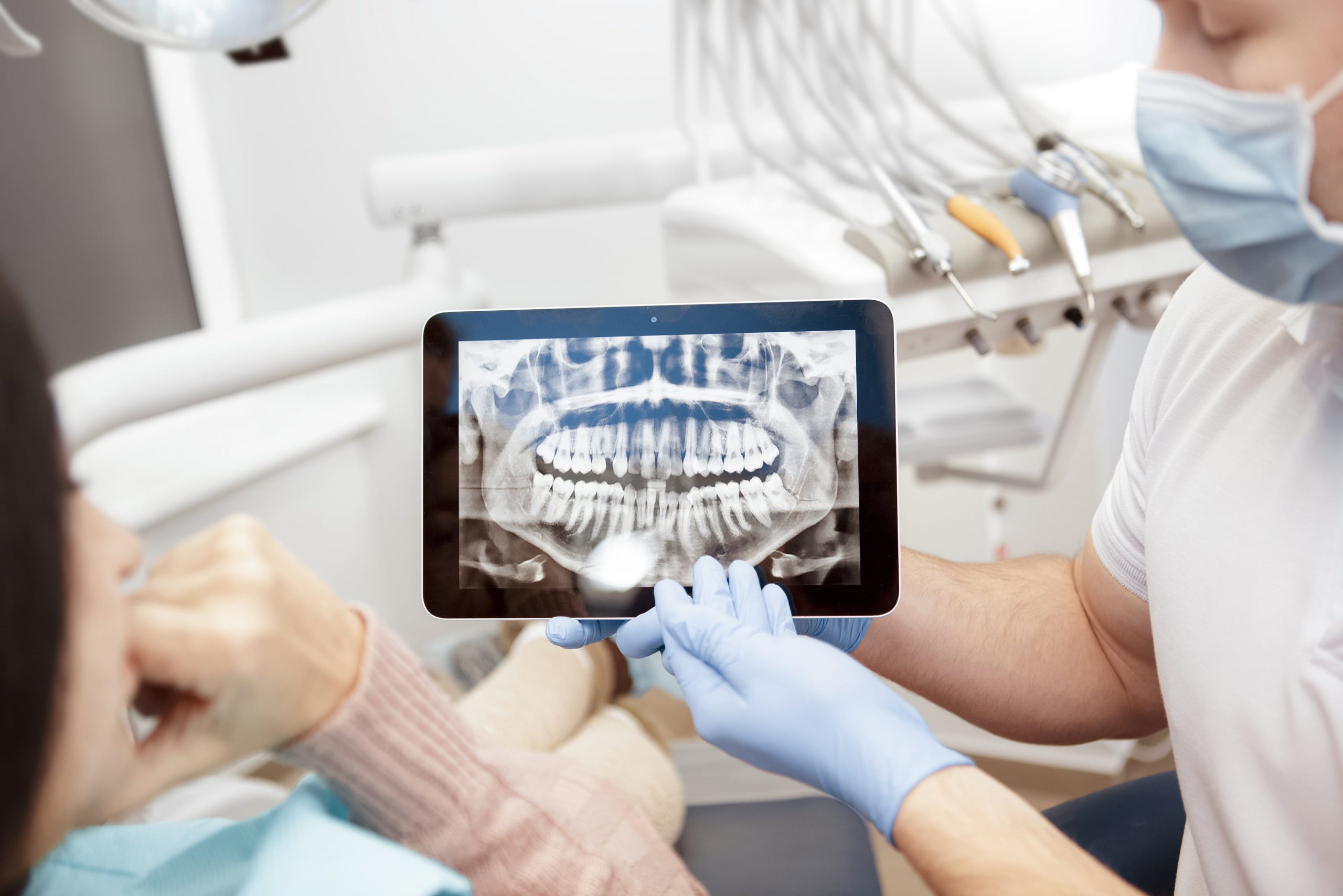
Tomography before dental implantation
All this data is transmitted to the computer, where it is processed in a special program. For implantation, conventional CT and multispiral (MSCT) can be performed (for example, for zygomatic implantation) – the latter can only be performed in specialized centers.
The removal of casts
These can be both traditional casts and 3D scans of the oral cavity. The resulting information is also uploaded to a computer program for further analysis.
3D visualization of the entire implantation process
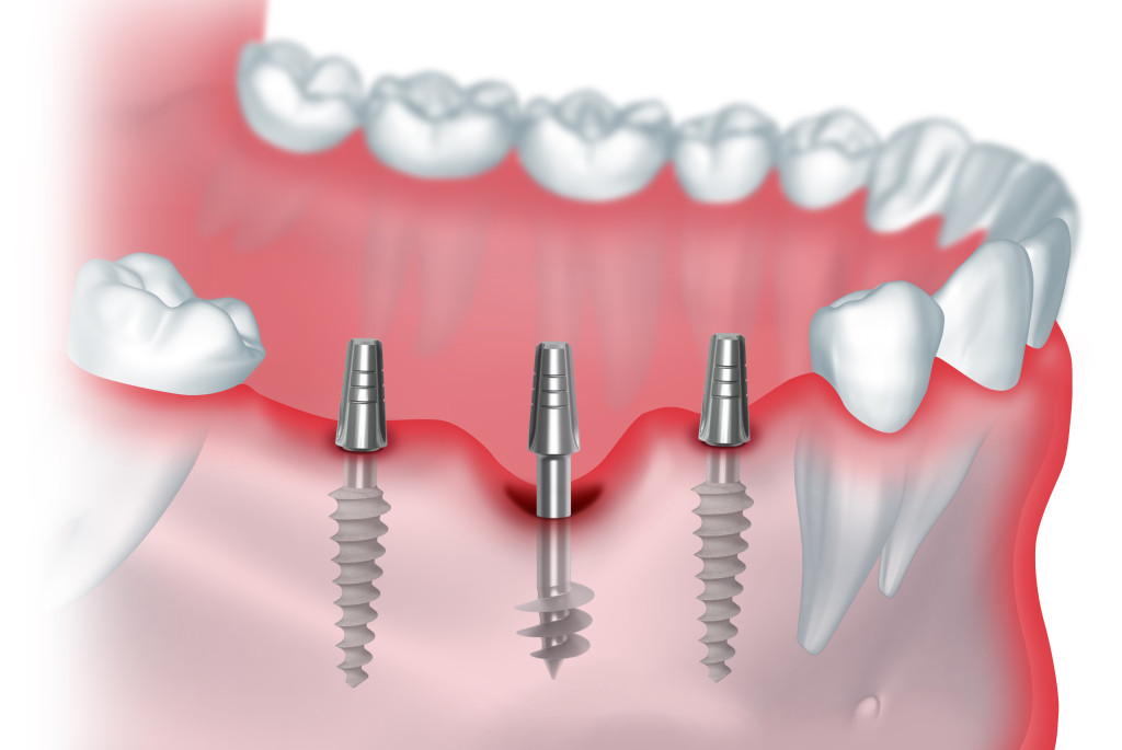
Based on the CT image, the program creates a three-dimensional virtual model of the jaw system. In it, the doctor can perform all the stages of implantation: removal of destroyed teeth, installation of implants in the appropriate places. The program helps you choose the right type and model of implants that will perfectly fit the patient. Together with the planning of dental implantation, the orthopedic doctor is working on the model of the future prosthesis.
This takes into account the structure of the patient’s jaw, the presence and location of natural teeth, as well as nerve endings, maxillary sinuses – all this allows you to avoid unpleasant medical errors. Patient participation is not required at this stage.
Fabrication of surgical templates
The next step is to create surgical templates on a 3D printer – special stencils made of transparent flexible plastic for installation on the gums. Guide bushings for drills are placed in a certain position so that the doctor installs the implants as planned in the program. The ready-made template is used only for a specific operation, because each patient is different. Its use eliminates the risk of error in conditions of lack of bone and guarantees the installation of structures in the intended place without fear of hitting the nerves or sinuses.
Surgical stage
After all the preparatory procedures, a surgical operation is performed using templates, which is based on a well-developed plan. Thanks to the templates, manipulations are performed minimally invasive, by puncturing and then screwing in the implant.
Repeated removal of casts for modeling the prosthesis
When the implants are installed, it is necessary to fix their location on the casts, as well as re-remove the bite parameters using special analyzers. Since doctors already have a computer model of the prosthesis, created at the stage of computer planning, it is not difficult to adjust it with the new information.
Manufacturing and installation of the prosthesis
In the laboratory, a plaster model of two jaws is created, on which the future prosthesis is modeled. It is also scanned and loaded into a special program. Plus, the dental technician must use CT data, as well as the results of the first stage of computer modeling – to create a more accurate model of the prosthesis.
When developing a prosthetic model in modern laboratories, robotic equipment is used at all stages.
Digital smile design for dental restoration
Visualization helps not only in implantation, but also in prosthetics. Digital Smile Design (DSD) technology – digital smile design-allows you to see and “try on” your new teeth before the start of implantation. This is possible thanks to computer modeling, which takes into account not only the functional side of dental restoration, but also the aesthetics of the face as a whole.
DSD is based on the principles of the “Golden ratio” and the harmonious ratio of the face and teeth. The program evaluates the proportions of a real patient’s face and selects the most natural and beautiful teeth that match their shape and color. As a result, the patient receives not only a beautiful smile, but also a complete transformation of the face due to the effect of dental face lifting.
It is important that template solutions are not applicable here: the program processes unique CT, photometry, and intraoral scanning data. Modeling a future smile takes into account important bite parameters and anatomical features.
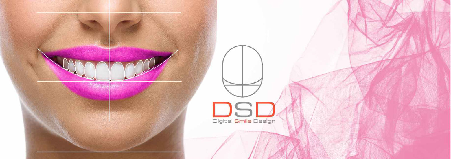
DSD creates beautiful prosthetics of any length, single crowns supported on implants, and veneers.
Advantages of 3D planning
The 3D modeling technique significantly saves the patient’s time and reduces possible risks. This is an innovative, high-tech and completely safe method of dental implantation for the patient, which has many advantages:
- safety: accurate placement of implants even in complex cases of atrophy, there is no risk of damage to the nerves in the lower jaw and the maxillary sinus shell located above the upper jaw,
- visualization: as for the doctor and the patient, the physician can clearly demonstrate to the patient all the stages of implant insertion, as well as calculate all the parameters of the operation
- predictability: what will be the result, we can say even after the creation of the model
- the reduction of the human factor: experiments confirm that in comparison with the installation according to the classical method of 3D-implantation is a better approach, avoiding erroneous actions,
- minimal trauma surgery.
The next stage of 3D implantation development is real-time visualization
The described 3D implantation approach is called static navigation. In recent years, developments in the field of dynamic navigation have appeared in dentistry. We are talking about X-Guide technology, a platform that combines programs and equipment for implantation and visualization in real time. In other words, in addition to the 3D implantation capabilities described above, the right to monitor the implantation of implants on the monitor is added at the surgical stage and adjust the operation in the course.
this technology is still being tested – and then only in one dental center. Due to the high cost of the complex and the lack of qualified personnel, its widespread use is currently difficult to imagine, but in European countries and the United States, X-Guide is gaining more and more supporters.
Digital technologies in dentistry are not the fantasies of futurists, but technologies implemented in the work of specialists, which are gradually becoming the standard in this field and even common practice.