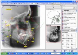
After analyzing TRG, calculating jaw models, and studying the face, an important part of orthodontic treatment is its planning and control. A variety of sophisticated methods for evaluating results have been created by several schools and allow the orthodontist to be confident in their diagnosis. Although, applying some research can be unnecessarily difficult and time-consuming. The current level of computerization of dental clinics creates the basis for the development of software designed for the analysis of digital images in orthodontics. This in turn, on the one hand, improves the quality and reliability of the diagnosis, and on the other – reduces the cost of material, effort and time. These goals determined the development of a whole group of onyx programs for image research.
More than 80 survey methods for various types of 2D and 3D images are available in ONYX-Ceph3. This library is constantly updated. The survey process is simplified to the placement of landmarks on the image required for this survey. Identification of landmarks is facilitated by graphical tools and other features of the program. Up to 1000 test results can be saved for each patient. You can access this data at any time and print it if necessary. It is also possible to exchange data over the network with technical experts and colleagues. Software updates for registered users are free of charge and available over the Internet.
Table of Contents
Competence
The onyx Ceph 3 ® software concept has been developed and developed in collaboration with highly qualified orthodontists, maxillofacial surgeons, dentists, and programmers. The structure and algorithm of the database is determined by the main course of action when generating images for diagnostic purposes. All professional methods and tools for orthodontic diagnostics are presented in the reference file. In addition, the great advantages are the flexible interface of the program and its easy adaptation directly to each user without interfering with the structure. With drivers and standard interfaces, Onyx Ceph 3 ® is compatible with all commonly used digital image sources. Data transfer from control systems is supported by a user-friendly interface. Each onyx ceph3 ® installation creates a client from a virtual Onyx network that can participate in online or offline data exchange within laboratory services, training sessions, or projects run by multiple organizations.
Profitability
To use Onyx Ceph 3 ® in their practice, the user does not even need to purchase the software, but simply pay an annual maintenance fee (rent), which includes a network license for a maximum of five workstations, regular program updates, and access to all available options at no additional cost or hidden costs. If in the process of using you still want to buy, then on favorable terms.
Friendliness
The user can always work with the current version of the program. In a timely manner, before the license expires, the program automatically offers its renewal. If the license is not renewed, all existing results will remain fully available, but new research will not be available.
Onyx Ceph 3° is supported by phone, email, and mail.
Functionality
The Onyx Ceph 3 ® software includes components for examining all major types of orthodontic images (lateral and frontal telerentgenograms, hand radiographs, jaw models, and facial photographs). For an additional fee, a 3D module is also available, where you can upload and process already three-dimensional images, such as jaw models, CT scans, and three-dimensional photography. All results belonging to the selected patient can be processed using the same software tool sets and approaches to solving the tasks set. The image processing process is simplified and automated to save time.
Onyx Ceph 3 ® is compatible with all digital image input sources. It is also possible to integrate with the clinic management system. The results of the study can be transmitted via the Internet to colleagues or experts.
In addition to local network capabilities, a special client structure allows online or offline sending of images within the onyx virtual network. It is possible to evaluate images in computers that are not integrated into the network and update these images in the main database, as well as transfer images to colleagues or to diagnostic laboratories, insurance companies, experts or other clinics.
You can save time by creating your own printed forms for various documents, such as contracts, informed consent, and others.
The program has its own presentation module, an analog of PowerPoint from the familiar Office package.
To demonstrate devices and clinical cases to patients, and to motivate them to orthodontic treatment, the program has its own media presentation module, which is configured individually for each user according to their tastes.
Image source
The onyx ceph 3 ® diagnostic software can be used independently of a specific image source with all available digital images that are relevant to a specific patient. For example, snapshots from the image database.
In addition to the special onyx IDS image input module, you can also use other digital sources (scanners, digital cameras, x-ray digital devices, clipboard, etc
Diagnostic method
 Onyx uses various image analysis techniques for research. These techniques are grouped according to the type of image being studied.
The manufacturer presents methods for studying the following types of images:
- Lateral TRG
- Front TRG
- Optg
- TRG brush
- Models of temporary occlusion
- Replacement bite models
- Models of permanent dentition
- Profile photo
- FAS photo
- Photo of a smile
Using the PVL option, you can create additional methods for analyzing existing or other types of images.
Treatment modeling
The studied images can be transferred to the planning module, which allows you to simulate treatment and create clear visual images of the expected treatment results (photo, TRG outline).
Using a two-dimensional image, surgical or orthodontic treatment can be simulated by segmentation, movement, and rotation of various user-selected areas.
The results of the selected analysis are processed during the simulation and can be saved as modified studies in the patient profile file, which allows you to compare, overlay images and use them with all available functions in Onyx Ceph3 ® .
Documentation
The documentation provides a standard guide to the idea of reference and a reference guide to all diagnostic techniques, applied landmarks, and obtained linear and angular results. Unfortunately, all documentation is currently available only in English or German.
In addition, on-line assistance is provided in the reference book of all established analysis techniques with links to source materials, which are also listed in the Help menu and provide a professional reference book with systematic and detailed explanations.
Before making my choice in favor of Onyx Ceph3 ® , I studied about a dozen programs with similar features and capabilities. I did not stop getting acquainted with the diagnostic software after this choice, periodically receiving demo versions from different manufacturers in one way or another. While my opinion does not change, today the Onyx Ceph3 ® software is the optimal price-quality ratio. This is actually the only foreign product with high-quality support, which is constantly updated and developed, and what is very important for many doctors, it is in their native Russian. And believe me, if you find any error in the translation and report it, it will be fixed very quickly.
So, each of you has the opportunity to make your own choice or leave everything as it is. And this is at best again to take up pencils, protractor and tracing paper, well, we will not even think about the worst, because we should all strive to improve our skills not for the sake of our ambitions or any other selfish motives, but first of all for the sake of patients who come to us with the hope of finding a beautiful smile without complications.

