ImagingPlus ™
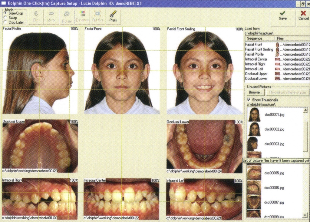
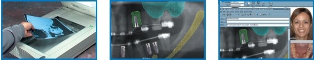
The main module of the Dolphin Imaging software. Allows you to manage photos, images, and x-rays. Create a presentation of your treatment plan in an accessible way for the patient using powerful and intuitive tools.
- organize patient photos in the desired sequence inside a standard template, and use a wide toolbar for image editing;
- import x-ray images and other images using the TWAIN standard, full integration with digital x-ray machines;
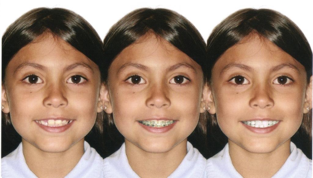
- orthodontic and surgical modeling: quickly changing the contours of the patient’s profile to demonstrate the predicted result of treatment, modeling of frontal images, detailed drawing tools;
- creating automatic cephalometric markup;
- compare images step by step in real time;
- ability to print x-ray images in 1:1 scale;
- creating PowerPoint presentations;
- improving communication with the patient.
Quickly get a series of patient photos
Modeling of treatment: before, at the stage, after.
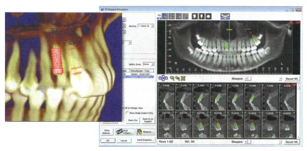
Implanner ™
A multi-functional and easy-to-use software module for planning implants directly on the patient’s x-ray image.
- a set of tools for improving the quality of x-ray images;
- modeling the spatial position of the implant in the bone;
- performing high-precision measurements;
- planning operations to improve the condition of the supporting bone.
- Scan the x-ray image or upload a digital equivalent.
- Modeling the position of the implant, drawing the required landmarks
- Print out the created treatment model and send it by e-mail to your colleagues or patient.
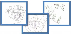
CephTracing
Cephalometric analysis allows you to quickly and accurately create cephalometric markings and perform the corresponding analysis of telerentgenograms, saving time and increasing the efficiency of Your work.
- import TRG from existing files, get images using a scanner, digital x-ray, extract from three-dimensional CT images;
- use in orthodontic and surgical practices: fully customizable front and side analysis by Ricketts, McNamara, Steiner, Roth and more than 400 others;
- analysis of dental arches, modeling patterns of various cephalometric structures;
- combining TRG of different stages of treatment, applying cephalometric contour lines to the patient’s profile photo;
- powerful image quality enhancement system for revealing hard-to-see structures.

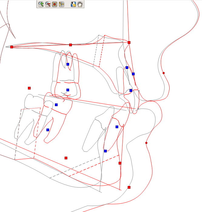
The combination of TRG and photographs of the patient profile
The combination of TRG (lateral ,frontal analysis)
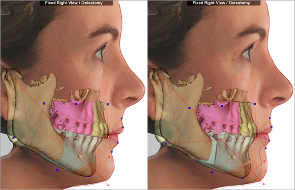
Treatment Simulation
The treatment Simulation software module includes interactive tools for high-precision diagnostics, planning, and creating presentations of clinical cases in a side projection.
- orthodontics: movement, rotation of incisors, molars, autorotation of the lower jaw, work with the dental arch, occlusion;
- Growth Forecast-forecast and modeling of growth based on TRG markings or patient photos;
- development of a treatment plan based on standard and individualized cephalometric analysis;
- VTO Wizards and Analyses-orthodontic and surgical treatment planning module (AEO – Roth/Ricketts, Arnett, McLaughlin) modeling treatment according to a specific cephalometric goal;
- orthognathic surgery (LeFort, BSO, osteotomy, genioplasty, zygomatic bone grafting), soft tissue contour correction tools based on changes in bone structures;
- simple intuitive use of the module with the mouse.
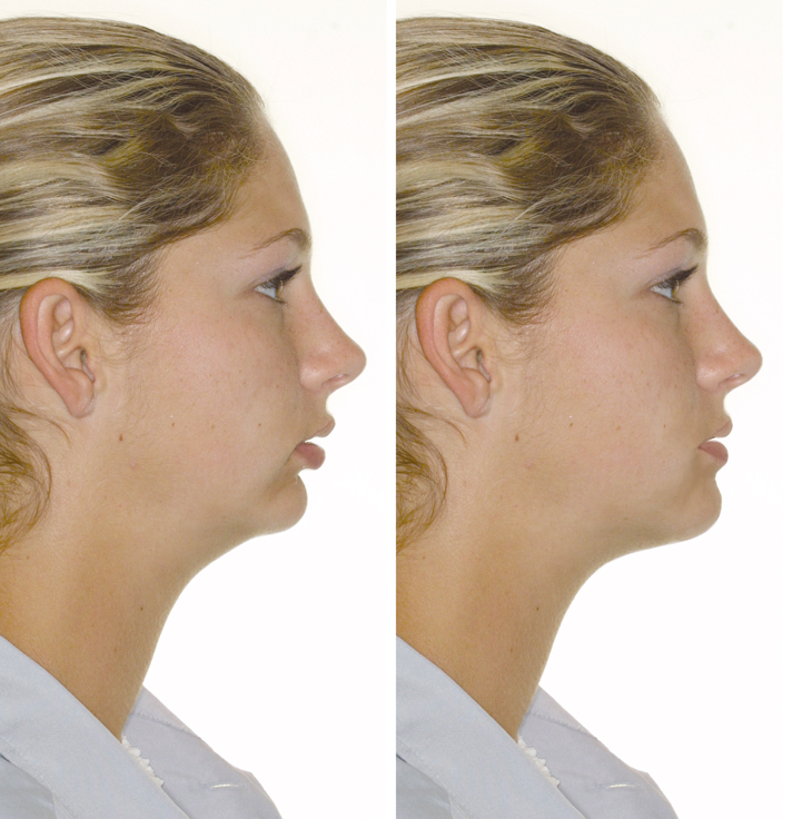
Simulation of surgical treatment
Planning of surgical treatment
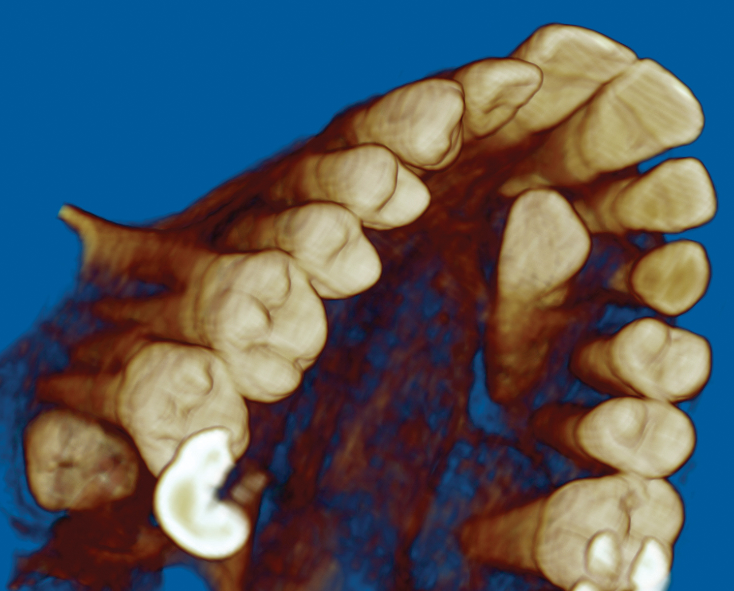
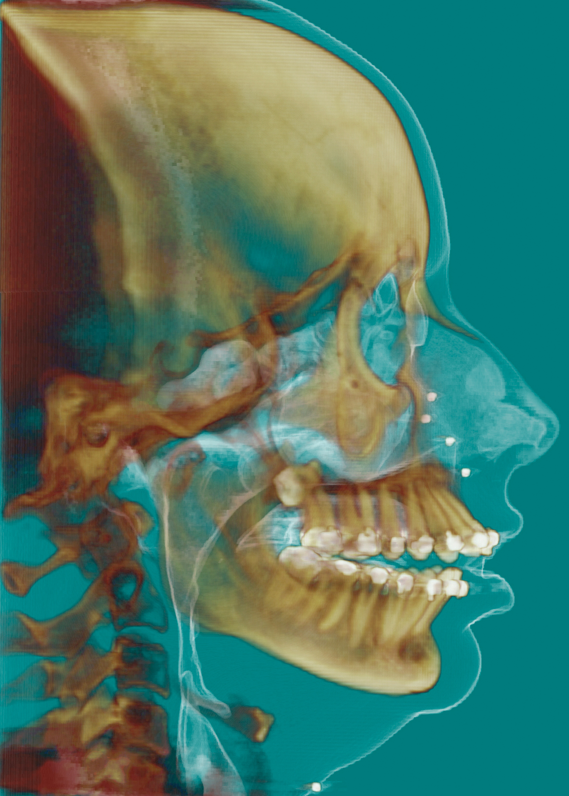
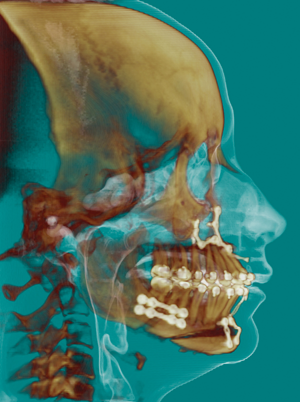
Dolphin 3D
Dolphin 3D three-dimensional software is widely used in research institutes, universities, and clinics around the world. Dolphin 3D is equipped with tools for three-dimensional visualization and analysis of large data sets that make working with 3D images as simple and efficient as possible! Allows you to view and analyze 3D images obtained using CT, MRI, and 3D cameras.
- image rotation, changing the level of tissue density;
- three-plane analysis of air spaces;
- working with CT slices in Multiple PlanarView (MPV) ;
- ability to combine two 3D images into one;
- superimposing a 2D photo of the patient’s face on a 3D image of the skull;
- 3D marker of the mandibular nerve;
- getting TRG and orthopantomogram from 3D;
- 3D and 2D measurements: distance, angle, area;
- changing the radiopacity to highlight individual anatomical structures;
- 3D modeling of dental implants + planning of sinus lifting and lifting of the nasal cavity floor-new in version 11.5!;
- detailed view and analysis of the structures of the TMJ;
- creating a virtual surgical guide templates.
Analysis of the situation of impacted tooth from any angle
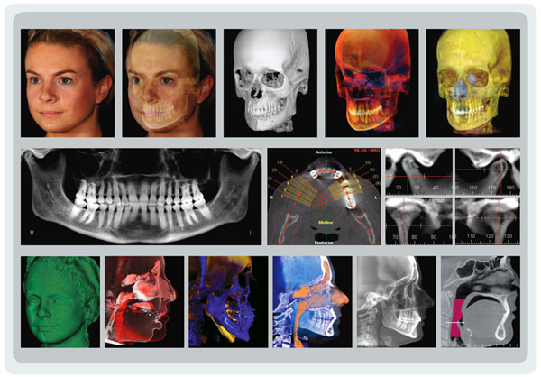
Presentation of treatment results: before and after surgery
3D modeling of the implant position
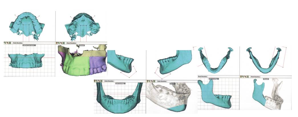
3D Surgery
3D Surgery – the latest module for planning surgical operations, animated demonstration of skeletal and soft tissue changes in the maxillofacial region using 3D images of spiral and cone-beam tomography.
- step-by-step actions: from initial analysis to modeling the final sprint;
- selection of individual fragments of the maxillofacial region;
- drawing cephalometric analysis points on 3D CT;
- occlusion fixation-combining 3D CT with 3D STL model of the jaw;
- marking osteotomy lines, moving fragments to visualize the outcome of the intervention;
- the possibility of an animated demonstration of the results of treatment .
Planning of osteotomy according to the clinical diagnosis.
Demonstration of the surgical plan: before and after treatment
3D splint modeling for orthognathic operations

Aquarium
Aquarium is a collection of high-quality, professionally executed three-dimensional multimedia materials covering various topics. An interactive module with a colorful interface allows you to visually demonstrate the patient’s diagnostic results, the strategy of orthodontic or surgical treatment, and show training videos on hygiene and the rules for using orthodontic equipment.
- an extensive video library of materials on various topics – each topic is presented in a series of presentations organized according to the diagnosis and treatment option;
- combining video, 3D animation of the treatment plan, and before/after clinical photos;
- accompanying the video with comments from the attending doctor;
- create a personalized playlist of materials that can be sent by e-mail to colleagues and relatives of the patient.